
Dental RX
Our Technology
Latest-generation digital radiology equipment. High-precision diagnostics with the lowest possible radiation dose.
Core technology
3D Cone Beam Computed Tomography (CBCT)
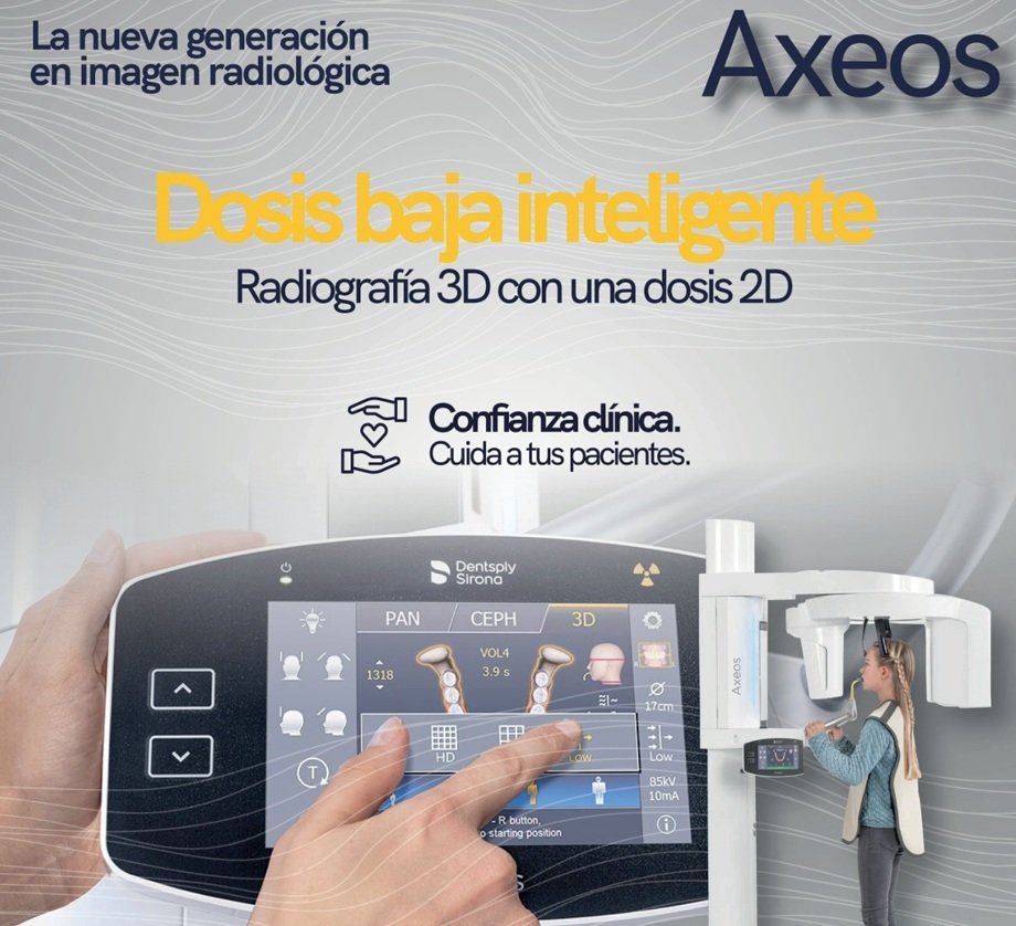
CBCT (Cone Beam Computed Tomography) is the gold standard in three-dimensional dental diagnostics. Unlike conventional medical CT, dental CBCT uses a cone-shaped X-ray beam that captures a complete volumetric region of the oral cavity in a single rotation, with a radiation dose up to 10 times lower.
The result is a high-resolution 3D reconstruction that allows the dentist to assess bone density with millimetric precision, locate critical anatomical structures (inferior alveolar nerve, maxillary sinus, nasal fossa), detect subclinical periapical lesions and accurately measure available bone volume for implants.
Our latest-generation CBCT equipment produces images with voxels as small as 0.08 mm, guaranteeing the resolution needed for the most demanding cases: advanced implant planning, complex endodontics, orthognathic surgery and TMJ assessment.
Technical specifications
· Voxel resolution: up to 0.08 mm
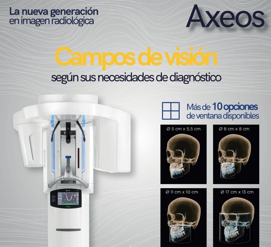
· Fields of view (FOV): 5×5 cm to 18×16 cm
· Exposure time: 14–26 seconds
· Effective dose: 40–150 μSv (depending on FOV)
· Delivery format: DICOM + JPG/PNG
· Multiplanar reconstruction: axial, coronal, sagittal
· Compatible with: Simplant, Nobel Clinician, coDiagnostiX, Implant Studio, Exoplan
Main clinical applications
Implant planning · Assessment of impacted teeth · Diagnosis of periapical lesions · Complex endodontics · TMJ · Orthodontics (3D cephalometry) · Orthognathic surgery
All our diagnostic equipment
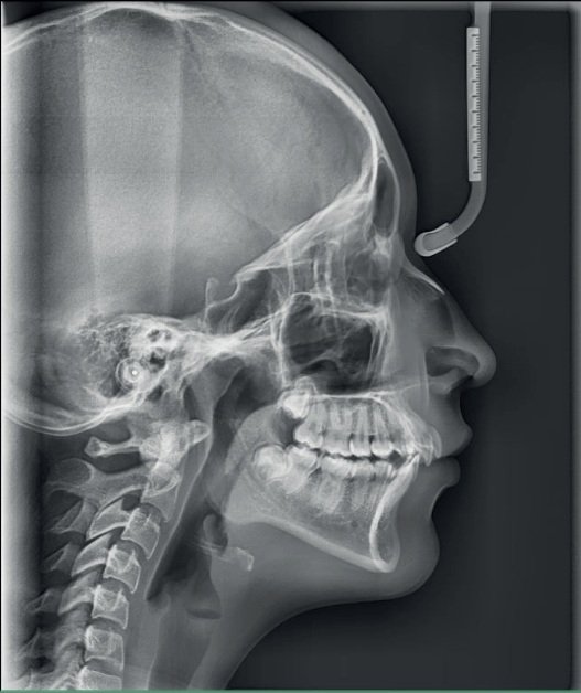
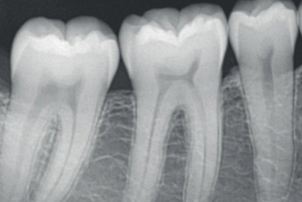
Each imaging modality has specific indications. Our oral radiology technicians help you choose the right study for each clinical case.
Digital workflow — from the study to your software
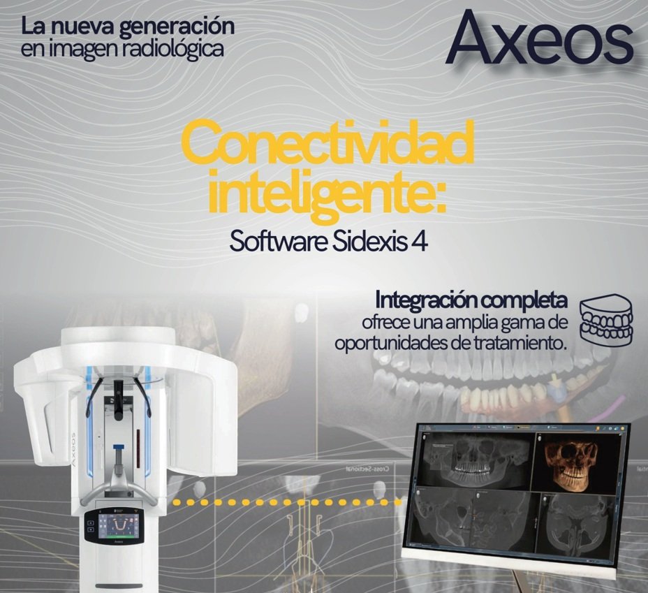
Our goal is for you to receive the image ready to import into your planning software, with no intermediate steps.
01
Capture
The patient is positioned in the equipment. The study takes between 14 and 26 seconds. No prior preparation is required.
02
Reconstruction
The software automatically reconstructs the 3D volumes in every plane: axial, coronal, sagittal and 3D rendering.
03
Report
Our specialist generates a structured radiology report with the findings and clinical recommendations.
04
Delivery
Receive the DICOM file plus a PDF report by email or USB the same day as the study — ready to import into your software.